
Photo by Ehsan Rezabeigi
Using metallic implants to replace a piece of bone damaged by disease or an accident can lead to serious complications, including infection and rejection by the body. A Concordia University postdoctoral fellow is working on a better way, using a lightweight implant made of glass and polymer foam that promotes bone regrowth and healing. These flower-like structures, pictured here, are made of a non-toxic and biodegradable plastic (polylactic acid; PLA) which is growing on the surface of a tiny bioglass particle with a diameter 10 times smaller than that of a human hair. These flower-like petals enable thousands of bone cells to replicate, while the bioglass particle gradually decomposes and provides these cells with the "food" needed to grow, including calcium and silicon. The result is a faster and better healing process for broken bones. Eventually, this implant will be completely resorbed into the body and replaced by new, natural bone.
Jury Prize
People’s Choice Award

Photo by Kate Donaleshen and James Robinson
A small parasitic wasp (Megastigmus spermotrophus) is one of many destructive insects to attack Douglas-fir trees, a major economic driver in western Canada. The insect lays its eggs in the seeds of the tree, where the larvae hatch and consume the content of each seed. Since there are no external signs of damage until the adult emerges from the seed up to five years later, X-rays are used to identify infected seeds. Researchers are using these images to study seed size, seed location within the cone and other traits to identify host characteristics that influence seed preference by these parasites. This image shows how a few Douglas-fir cones can support a high number of Megastigmus larvae, but also produce empty seed, and seed with developing plant embryos. The research reveals direct impacts on individual tree fitness—information that will prove useful in forest management practices.
Jury Prize

Photo by Simon Octavio Valdez Juarez
This yellow warbler is quite the long distance flyer, the coloured band seen on his leg is a testament to the species's migratory endurance. I banded this bird in January 2012, next to a tree dividing two chili-pepper fields in Jalisco, Mexico, where he hunted for spiders and caterpillars (as seen here). In April he migrated north and I lost track of him, but the following year he was back in the same tree. Feather analysis revealed that his breeding grounds were in the Yukon or the Northwest Territories, Canada. This picture was taken in January 2015. In the time between the banding and this picture, this tiny, nine gram bird migrated from Canada to Mexico four times, an impressive 30,000 km of travel. This yellow warbler is one of 200 being studied to understand how agriculture affects the ecology and survival of migratory birds.
Jury Prize

Photo by Youcef Bioud
The tightly packed stems in this image may look like a field of wheat, but they’re actually nanothreads of indium phosphide. The process for producing them is fairly simple: the compound is placed in a solution of hydrofluoric acid, through which an electrical current is then passed, and the filaments form quite naturally. The resulting three-dimensional structure is of particular interest to the photovoltaic-cell industry, because of the larger surface area that it provides for capturing the photons emitted by the sun. In fact, can sunlight can be absorbed both at this structure’s surface and below it, thus providing an even more effective way of harvesting solar energy. Each filament has a diameter of 20 to 200 nanometres.
Jury Prize

Photo by Chérif Matta
An astute biochemist will recognize these pairs of guanine-cytosine nucleic bases at first glance. This configuration of molecules may be familiar, but this image of them still represents a first, because the bonds between the atomic nuclei have not been drawn: they have been calculated by quantum methods. What you are looking at is actually a topographic map of the density of the electrons of each of the atoms that share this space. This density is illustrated by the small lines converging on the nuclei, and it is the interaction of these fields of potentiality that causes the optimal bond paths of electron density connecting the atoms to emerge.
Jury Prize

Photo by Émilie Peco
Here we are at the heart of the nervous system of a Drosophila fly larva, which functions on the same basic principles as the nervous systems of mammals, including humans. To the right are three pairs of astrocytes (white) delicately wrapped around longitudinal neuronal pathways (blue). Other astrocytes are present, but were not revealed by the marker used to capture this image. These glial cells, with their complex ramifications, are essential to the functioning of the central nervous system, but many of their roles have yet to be discovered. It is clear, however, that astrocytes are not distributed at random: they always cover the same neural areas. Scientists are still trying to understand the mechanisms that show them the way.
Jury Prize

Photo by François Rheault
We all have some vague idea of what grey matter is: the tissue at the surface of the brain, with its familiar folds. But we are less familiar with the white matter that lies beneath. This white matter consists of a multitude of networks of axons, which are extensions of the neurons and constitute the brain’s “wiring”. These axons are sheathed in myelin, a white substance that facilitates the transmission of electrochemical information. The organization of these networks of axons is the same in all brains, though each has its distinctive traits. Using medical imaging technology and powerful software, scientists can now dissect a brain virtually, so that only its main neural highways remain.
People’s Choice Award

Photo by Jill Brooks
Great hammerhead sharks are among the most endangered species, driven largely by Asian demand for shark fin soup and accidental bycatch from commercial fishing. Global conservation efforts are underway but they rely on knowing where these fish travel, which habitats they prefer, and how often they leave sanctuaries and swim into commercial fishing zones. In this picture, Carleton University MSc student Jill Brooks is free-diving to shoot a dart tag into the back of a shark—one of 32 tagged with an acoustic transmitter to track their movements throughout the Bahamas and the southeast coast of the United States. Data from the transmitters are sent to multiple receivers anchored to the ocean floor and shared with organizations around the world, including Canada's Ocean Tracking Network.

Photo by Daniel MacPhee
How uterine contractions during pregnancy and labour are controlled is still poorly understood. Yet, unravelling this mystery is vital if we hope to understand conditions such as preterm labour, which affects 5-10% of human pregnancies in North America and is responsible for 75-85% of neonatal deaths and morbidity. Problems during labour and birth also exist in large domestic animals with similar consequences. In this image of a labouring rat uterus, researchers used a high-intensity fluorescence microscope to illuminate a stream of uterine smooth muscle cells containing high levels of small heat shock protein B5. During labour, these "molecular chaperones" help produce muscle that generates coordinated and powerful contractions to ensure the timely delivery of a term fetus. This basic research could one day help physicians and veterinarians predict when labour processes could become abnormal—and in the process help protect the health and lives of both newborns and mothers.

Photo by Timothy Gibson
Nunavut is home to the billion year-old, Angmaat geologic formation. Now exposed high on the coasts of Baffin Island, this ruggedly eroded rock layer once accumulated on the floor of ancient seas. These 500 metres of ancient marine sediment can be studied with the latest geochemical techniques to learn about changes in ocean chemistry, the formation of the vanished supercontinent Rodinia and new clues to the pace of evolution of very early life. New geological dating for these rocks has revealed that the first known sexually producing organism—a primitive multicellular red alga called Bangiomorpha pubescens —emerged nearly 200 million years later than previously thought and may be related to important changes in Earth's environment. The Precambrian Research Office and Publican Society at McGill University are conducting ongoing investigations of the Canadian Arctic's rich endowment of ancient rock to reveal Earth's past climates and environments.

Photo by Émilie Bouchard
Toxoplasma gondii is a parasite that infects warm-blooded animals, including birds and mammals. In most cases, the infection causes few symptoms but can lead to neurological, ocular, and reproductive problems, especially if the immune system is compromised or if a mammal contracts the parasite while pregnant. Humans can also become infected through contact with soil contaminated by the feces of cats or through eating infected meat. Unexpectedly, the bug is appearing in Arctic fox in Nunavut, where the main carriers, domestic and wild cats, are practically non-existent. Researchers are collecting and testing blood samples from live-trapped adult and juvenile Arctic foxes to determine if the parasite is transmitted from mom to baby via the placenta. This research will provide important information about the health effects in wildlife and how Arctic people become exposed.

Photo by Phil Angel
Climate change threatens biodiversity, forcing species to move to new areas and others to disappear from the hottest areas. Butterfly biology depends strongly on temperature, and can act as a proverbial "canary in the coal mine", providing an early warning signal for climate change impacts on other species. Butterflies, and other pollinators (e.g. bumblebees) provide irreplaceable ecosystem services in agricultural landscapes and natural ecosystems. At the University of Ottawa, macroecologists have worked to collect huge numbers of butterfly observations, building on biodiversity baseline data from over a century of climate and habitat changes. Long-term data such as this have been used to demonstrate rapid pollinator declines in many parts of Canada. Pictured here is the Viceroy butterfly (Limenitis archippus), famous for its mimetic relationship with the Monarch butterfly (Danaus plexippus), and captured during sampling at a field site near Ottawa.

Photo by Mais Aljunaidy
What happens in the womb can have a powerful impact on a person's future health. Some 20% of pregnancies are affected by complications such as pre-eclampsia or poor fetal growth (e.g. low birth weight) that increase the chances of developing cardiovascular disease in adult life. However, the mechanisms underlying these changes are not known. Researchers at the University of Alberta are studying how harmful compounds released by the placenta during complicated pregnancies can cause the mother's blood vessels to no longer work properly, leading to increased blood pressure and altering the cardiovascular development of her offspring. This image shows the most prevalent cells in the heart—cardiomyocytes and fibroblast cells—forming the cardiac wall in fetal rats.

Photo by Wenge Jiang and Marc D. McKee
Nature inspires and instructs us to rationally design and build novel biomaterials that are useful to humankind. At McGill University, researchers are growing hierarchically organized biomaterials with spiralling suprastructures resembling those found in nature, including in snail shells and seashells, and in pathological "ear rocks" (otoconia), occurring in disease of the human inner ear. Working initially at the nanoscale, the team created complex handed (chiral), spiralling structures made of calcium carbonate mineral—the building block for the skeletons and shells of many marine and terrestrial organisms. To do this, they added a chiral acidic amino acid (glutamic acid). This photo shows six distinct interlacing coiled calcium carbonate platelet domains assembled into left-handed clockwise spiralling suprastructure arising from an unstructured centre core area (pink). The findings provide valuable insight into normal biomineralization processes, pathological biomineralization involving otoconia, and novel synthesis methods for next-generation materials.

Photo by Rebecca Hay
Chronic stress can contribute to multiple health problems, including depression, weight gain, and heart and liver disease. Researchers at Carleton University are studying how stress interacts with the gut hormone ghrelin to affect our health. Ghrelin—dubbed the "hunger hormone"—helps manage stress, weight gain and appetite. In this image, a liver was treated with the stress hormone corticosterone. The red represents fat deposits and the pink/blue wave through the image is scar tissue. The pattern of the scar tissue indicates a cirrhotic liver in the end stages of liver disease. This research found liver damage was most extensive in situations of both chronic stress and the absence of ghrelin signalling. The findings may have significant research and clinical implications related to obesity, diabetes and other chronic diseases.

Photo by Anastassia Voronova
The brain is a complex network of cells that forms during pregnancy as the embryo grows into a fully formed baby. To develop properly, the brain needs stem cells, including one type called cortical precursor cells. Found in the cortex—arguably one of the most important parts of the human brain as it is involved in highly complex processes like thought, memory, perception and action —these cells contain the intricate instructions necessary to produce a healthy functioning brain. These cells go through a process called differentiation in which they become one of three "neuronal" cell types. This image of a healthy, newborn, mouse brain shows all the cells arising from cortical precursors. We can track these cells because they contain a gene that produces a red-glowing protein. By comparing this healthy specimen with other brains, we can examine the effect of external factors on brain development and the link to disorders such as autism spectrum disorder and microcephaly.

Photo by Ashley Chin
Honeycomb patterns exist everywhere in nature, from bee hives to turtle shells. But did you know they can also be found inside the ovaries of the common fruit fly, Drosophila melanogaster? These unwelcomed household pests surprisingly share 75 percent of the genes that cause human diseases, making them an invaluable organism in biological research. This image shows developing ovaries that have been genetically modified to study cell polarity. The loss of this structural property is often a precondition and hallmark of cancer. In this image, a green dye was used to label the regions where a particular polarity gene, exhibiting honeycomb pattern, has been deactivated. This side-by-side comparison to the normal red regions is useful for identifying a gene that may be involved in controlling cell polarity, crucial knowledge for unraveling the origins of cancer.

Photo by Melanie Clapham
Seeing these two young subadult brown bears play fighting in Knight Inlet, BC was an unexpected sight. Such play is common among bears of the same family, but has rarely been recorded between non-littermates. In this picture, the older bear on the right is uncharacteristically taking a submissive role, as indicated by his puckered-lip and ear positioning. The younger male attempts to dominate the older bear, displaying partially-flattened ears and an open mouth with wide eyes. This University of Victoria/Brown Bear Research Network-led research on social play in brown bears forms part of a larger study on their social behaviour. The research challenges our current perception of bear behaviour, with important implications for bear conservation and public understanding.

Photo by Sara Al-Habyan
Breast cancer lethality is often caused by cancer cells colonizing distant body parts, such as the bones, lung and liver, to name a few. To limit disease progression and develop novel therapeutic targets for cancer patients, it is vital to study how cancer cells migrate from the organ of origin to other organs. At McGill University, researchers colored cancer cells with different fluorescent dyes, including red, blue and yellow. This allows visual tracking of cells in action, and how exactly they reach distant organs. Here, a mixture of these cells is plated on a glass surface with nuclei colored in grey.

Photo by Daryan Chitsaz
Amyotrophic lateral sclerosis (ALS) and multiple sclerosis (MS) are neurodegenerative diseases that attack the body's nerves, which connect to muscles. Researchers are working at the human embryonic stage to discover how hair-like nerve fibers, called axons, extend from the spinal cord to muscles throughout the body. They hope to understand how neurons and muscles communicate to support and guide each other's development, and promote nerve regeneration. This image shows a sheet of hundreds of red-labelled rat muscle precursor cells (young cells that mature into muscle cells) with a dense cluster of spinal motor neurons in green. The nuclei are stained blue. After just one day, the axons were stimulated to grow hundreds of times their original length and follow the contours of the muscle cells. The research is an important step to one day developing treatments for people who have suffered nerve injury or neurodegenerative diseases.

Photo by Yiming Xiao and Vladimir Fonov
Three-dimensional digital maps or "atlases" of the brain are dramatically transforming how doctors diagnose and treat brain disorders and injuries such as Alzheimer's disease, Parkinson's disease, autism, schizophrenia and drug addiction. But everyone's brain is different. Researchers have used medical imaging to come up with the average brain shape of a population. This image shows such a brain atlas made of 20 people's MRI scans, with the brain surface rendered in blue, blood vessels in yellow, and the rest of the head rendered artistically using a stylized colour map. With specialized medical image processing and visualization software tools developed at McGill University's NeuroImaging and Surgical Technologies Lab, the 20 images were deformed, averaged and rendered to create one sharp atlas as if from a single person. The average brain shape is important for detecting disease-related individual brain shape changes in computer-assisted diagnosis and disease staging.

Photo by Nabeel Ahmed
Look up … way up. In the 1960s, astronomers discovered the brightest phenomena in the universe. Quasars are celestial objects that form around supermassive black holes at the centre of almost every galaxy in the universe. Its blinding brightness comes from a glowing disc of fast spinning loose matter—called an accretion disc—that is slowly sucked into the black hole. Contributing to this powerful process are a quasar's fierce, cosmic winds (depicted in blue) that can reach speeds of up to 30,000 km/s (10% the speed of light!). Researchers at York University are studying how these winds may have inhibited a galaxy's ability to create new stars. The image displays an artist conception of what quasars may look like.

Photo by Masha Prager-Khoutorsky
The image shows a small area in the rat brain magnified 400 times. This heart shaped structure is a part of the mammalian brain (hypothalamus), responsible for "feeling" the hormonal and metabolic states of the body. Specialized nerve cells located in this region sense levels of salts, glucose, and hormones circulating in the blood and transmit this information to other parts of the brain. The nerve cells in this area also regulate the cardiovascular system, as well as sexual and reproductive behavior. The image illustrates how different cell types are organized in this vital yet poorly explored brain region. The study, led by Dr. Charles W. Bourque at McGill University, is part of a research program aiming to understand how the brain regulates blood pressure.

Photo by Glenn Tattersall and Cleo Leite, William Milsom, Colin Sanders, Viviana Cadena, Denis Andrade, Augusto Abe
Not all lizards, it seems, are cold-blooded all of the time. A Brock University-led study has discovered that a tegu lizard, a common pet that hails from South America, produces and retains its own body heating during the mating season. The research upends the accepted belief that ectothermic vertebrates—animals that apparently lack internal temperature control—are always the same temperature as the air around them. In this well publicized study, Brock researchers found the animal's body temperature rises up to 10 degrees Celsius higher than the ambient temperature due to large increases in metabolism and efficient control over blood flow. This infrared thermal image shows the metabolic process in action. By linking heat production to reproduction, the discovery advances a critical theory of how endotherms—vertebrates like us that regulate their own heat—evolved.

Photo by Mélanie Aubin and Valérie Lecomte
These ghostly silhouettes are the remains of diatoms, a kind of microalgae. The remarkably symmetrical blue specimen in the foreground is a species of Pleurosigma that is rarely found so intact in sediment samples from lakes in northern Quebec. This microstructure is actually the organism’s exoskeleton and is composed of silica. Diatoms are present in almost all aquatic environments and are therefore routinely found in sediment analyses. To perform these analyses, researchers dry the sediments, pass them through a screen, then sprinkle them onto a carbon adhesive tape in order to view them under an electron microscope. One application of such analyses is to study the distribution of metals in lake sediments.

Photo by François Chartier and Patrick Laprise
This is the inside of the embryo of a fruit fly a few hours before it hatches as a larva (a microscopic, wormlike organism). If it had to, it could breathe through the network of air-filled tubules shown in orange here, which carry oxygen throughout its body. Scientists study these tubules closely, because the mechanisms that govern their elongation and bifurcation in fruit flies have been retained throughout the course of evolution and operate in human beings as well, where their dysfunctions are the source of many pathologies. In other words, fruit flies can still lead to fruitful discoveries.

Photo by Dominique Claveau-Mallet
The tiny spheres in this image are pieces of slag—waste material from foundries where iron is extracted from ore. The good news is that this waste material could well be used to save North American lakes from invasions of cyanobacteria—blue-green algae that proliferate when the concentrations of dissolved phosphorus in lake water become too high. Slag contains lime, which reacts with the dissolved phosphorus, causing the precipitation of crystals of hydroxyapatite, the grainy mass in the upper part of the image. These crystals bind to the slag, where they continue growing, thus trapping more and more phosphorus. A clever idea for making water crystal clean.

Photo by Julie Constanzo, Martin Lepage, Benoit Paquette
This picture, produced by magnetic resonance imaging (MRI), shows the blood vessels of a rat’s brain, seen from above. The denser area to the upper right was originally healthy tissue but in this experiment was targeted with the kind of radiation used to treat brain tumours, in order to reproduce the side effects of this treatment. Although this radiation kills tumours, it also sometimes damages surrounding brain tissue. In a sort of “panic reaction”, the brain’s blood vessels then begin to grow in an uncontrolled fashion. But from now on, thanks to this innovative application of MRI, clinicians can effectively monitor such changes in these blood vessels, so that patients can be treated sooner with appropriate medications if necessary.

Photo by Julie Alix Denoncourt, Steve Charette, Richard Janvier
The protozoan Tetrahymena, of which only a fraction in blue is seen here, is a single-celled freshwater organism. In this image, it is returning from a hunt for the bacteria on which it feeds (the green spots). These bacteria are stored in a digestive vacuole along with the defecations produced by other protozoans (in orange). Once the protozoan has finished its digestion, it compacts the residue from its meal to form larger fecal balls (in red). But some bacteria that survive digestion may be trapped in these fecal balls. Once these balls have been evacuated, they will provide the bacteria with good protection against a decidedly hostile environment.

Photo by Yann Develle
Here we see the end of a mouse’s spinal cord, attached to the wall of a test tube. An arc of light, produced by a photographic technique called light painting, has just finished making this artistic representation of the rotulian reflex—the same one that makes your leg lift involuntarily when the doctor strikes the tendon below your kneecap. This stimulus (illustrated by the pink curve) is modulated in the spinal cord, then re-routed in the opposite direction by a motor neuron (following what is now the blue-violet curve), which causes the muscular reaction. The researchers who produced this picture are studying this neural circuit to improve the motor recovery of people who receive spinal-cord injuries.

Photo by Weimeng Ding and Ariel Wilson
The blue blob at the centre of this image is a macrophage—a type of immune-system cell—and the small yellow dots that it is in the process of devouring are nanoparticles of gold on the surface of a rat’s retina (the purple background). Clinicians have recently begun using gold nanoparticles to target cells and deliver medication to them in a localized manner. But this technique is not used very much in the eye, because it is such a complex, fragile organ. That is why this image of a retinal “street sweeper” in action, a phenomenon rarely captured on such a small scale, represents such a great piece of luck. It is a reassuring indication that gold nanoparticles used to deliver medicine to the eye will not accumulate there, because the macrophages will clean them up.

Photo by Pascal Dubé
To facilitate navigation on rivers, scientists model the currents carried by the tides. This image shows numerical predictions of the currents on the St. Lawrence River between Quebec City and Rimouski. Each colour represents a different direction in which the currents flow: red for north, light blue for south, green for east, and violet for west. The paler the colour, the faster the current. The thin green thread toward the top of the image represents the Saguenay River; the white area at one end represents the intense activity where it flows into the St. Lawrence. This model represents only a snapshot of a moment in the life of this river. In reality, its currents are shifting constantly; in a few hours, this colour picture will be completely different!

Photo by Maeva Giraudo, Magali Houde, Guillaume Cottin
This picture shows a member of the genus Daphnia, a very common type of freshwater zooplankton. It has a single eye and uses its antennae mainly to swim and to feed. Beneath its transparent carapace, the orange structures are hemoglobin proteins, which carry oxygen. This organism’s digestive tube is green because its diet contains a high proportion of microscopic algae. Below, a few eggs can be seen, which this individual may have produced without a mate to fertilize them, a process known as parthenogenesis. It is essential to study Daphnia and the effects of pollutants on their growth and reproduction, because they constitute the bottom of the food chain for thousands of species. In fact, if Daphnia were to disappear, the entire aquatic ecosystem would collapse.

Photo by Mathieu Lapointe
Every day, we throw our glass containers in the recycling bin. As of yet, there are very few innovative ways to recycle crushed glass. Thanks to a creative technique, however, recycled glass can now be reused in wastewater treatment processes. When glass fragments are submerged in a basin of wastewater with a binding polymer, they capture a fair share of contaminants. By pure chance, the glass fragments pictured formed a shimmering flower that had sunk to the bottom of the treatment basin like an anchor. This promising water-purification technique is already just as effective as the conventional technique of using sand extracted from the ground.

Photo by François-Joseph Lapointe
This is a social network for bacteria. To produce this image, the researcher shook hands with 1001 people with whom he crossed paths in the street. As he did so, at regular intervals he took samples of the micro-organisms on the palm of his hand, which grew more and more diverse. Then he analyzed the DNA from these samples and used visualization software to translate the resulting data, assigning a different colour to each sample. In this image, the strands connecting those bacteria that had a genetic similarity of at least 95% are in some cases so dense that they look like balls of yarn. Contrary to what many people think, most bacteria play a beneficial role in the human body, and this kind of experiment provides a better understanding of how they behave “in society”.

Photo by Andrée-Anne Marchand and Julie O'Shaughnessy
As people age, they sometimes develop osteoarthritis, a form of degeneration of the spinal column. In the above X-ray, the areas of degeneration in two superimposed lumbar vertebrae are shown in yellow and blue. The overlapping area, in green, shows how the spinal canal has narrowed, compressing the spinal cord. This condition, known as central spinal stenosis, causes loss of mobility and acute pain. Although this process is not reversible, specific types of physical training can improve patients’ quality of life while they are waiting for surgery.

Photo by Jolyane Meloche
Every cell in the human body contains myriad organelles called mitochondria that produce energy. But in doing so, these tiny power plants also slowly burn out the cells in which they are located—a slow, entirely natural, programmed process of cell death. Cancer cells, are an exception, however: they refuse to die. Instead, they deactivate their mitochondria and keep on multiplying. That is the case here for pulmonary arterial hypertension. In the cell shown here, the deactivated mitochondria have been marked with a fluorescent orange dye, while the cell’s nucleus is a ghostly blue. This marking technique makes it possible to determine whether deregulated (cancerous) cells respond to treatment and abandon their suicidal quest to live forever. Confocal microscope and mitochondrial membranes marked with dye.
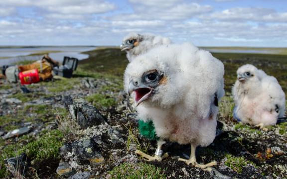
Photo by Alexandre Paiement
These chicks will soon grow into majestic peregrine falcons. In the centre, male number 220612893, 25 days old, shows off the identification band that will let researchers track him for the rest of his life. The green mark on his side means that he is the youngest member of his clutch. Researchers have studied him and his siblings closely ever since they were hatched, measuring their height and weight, taking blood samples, and so on, much to their mother’s displeasure! For over 30 years, adventurous scientists have been monitoring the population of peregrine falcons that nests on the cliffs of Rankin Inlet, in Nunavut, whose survival is threatened by environmental pollution.

Photo by Andrew Plowright
This map is a three-dimensional model of a neighbourhood in the town of Surrey, British Columbia. The image was created using a laser remote sensing device placed on an aircraft (lidar). Each coloured point indicates the position where a beam of light was reflected by the surface. Precise to the nearest centimetre, this technology creates highly detailed maps that provide a wealth of information. In this model, the data were collected to evaluate the health of the urban forest. This partially digitized image shows the number of trees, their height (seen in the yellow-to-red gradient) and their foliage density.

Photo by Tony Savard, Julie Barrette, Denise Chabot
The blue object in the foreground, against a background of green vegetable particles, is the bane of the canning industry: a Clostridium botulinum bacterium, notorious for the deadly toxin that it produces. Normally the members of this species are rod-shaped, but this one is in survival mode: the swollen end is a spore that it is forming—a copy of itself. This species is very hardy, but luckily, it can be destroyed simply by heating to high temperature. However, such heating also destroys certain vitamins, proteins and fibres that vegetables contain. Researchers are therefore looking for new methods of killing this interloper while preserving these nutrients.