
Photo by Rahaf Nafez Hussein
This image shows the degradation of an exposed film of copper in contrast with a thin protected layer that has not degraded—creating a picture resembling the blue waters and sandy shoreline of a beach. The small area at the top of the image has maintained its natural copper colour thanks to a protective coating. We investigated the ability of this biocompatible coating by laminating it over the surface of 100-ångstrom-thick evaporated copper on a glass substrate (the dark portion at the top of the image) and then exposing the sample to 100% relative humidity. This extreme level of humidity accelerated the corrosion of the exposed copper, forming this blue lace-like network, which preserved the copper’s conductivity. The laminated section of copper did not corrode as visibly, and this was reflected by better electrical conductivity, which shows that the biocompatible protective coating was successful. My research focuses on investigating the relationship between molecular structure and the physical properties of biocompatible films and next-generation materials. The aim is to create a library of eco-friendly protective materials to keep our planet safe and clean.

Photo by Antoine Durocher
Luming Fan
Patrizio Vena
Jeffrey M. Bergthorson
Marc Füri
Gilles Bourque
David May
Julien Sirois
Luming Fan
Patrizio Vena
Gilles Bourque
David May
Julien Sirois
Hydrogen alone burns without any carbon emissions, but it is intrinsically more unstable than current hydrocarbon fuels. Equipment can be damaged by using traditional combustion technology to burn high-hydrogen content fuels. This image shows a novel 3D-printed burner being used to stabilize high-hydrogen content flames into pure hydrogen. The burner relies on micromix technology—internal miniature fuel channels that are printed in the burner injector body to deliver fuel to localized sites. The five injectors are placed in a plus-sign shape to mimic the conditions of a hot gas turbine for the center flame. Then, we use non-intrusive laser diagnostics to investigate the combustion phenomena of these micromixed, high-hydrogen content flames and identify promising low-emission injector designs.

Photo by Ana Isabel Carrazco Quevedo
Nanoplastics (NPs) are found all over the world, and research that tackles this major environmental problem is urgently needed. This image shows NPs (yellow-green) in a water flea (Daphnia magna). We used confocal microscopy to locate the NPs with their intensity signal. D. magna are ideal for use with fluorescent-based microscopy techniques, because their bodies have blue autofluorescence. To achieve this image, we exposed D. magna neonates to 20 nm fluorescent polystyrene NPs (yellow-green) for 24 hours (acute toxicity). We can see the internalization of NPs in the gut, as well as some gut disruption (fragmentation) caused by NP uptake. NPs can also be seen in the antennae and claws, which D. magna use to navigate the water. The thoracic limbs are also stained with a yellow-green colour, indicating that NPs were retained as part of the filtration process.

Photo by Jean-Philippe Ayotte-Beaudet
Quebec is experiencing a remarkable boom in outdoor education. The benefits of this field have been widely documented, both in terms of contact with nature and on the cognitive, physical, psychological and social levels. However, new data collection tools are needed to assess learning. In December 2022, a team from the University of Sherbrooke spent a day observing first graders in Ms. Marie-Line's class at the Académie des Sacrés-Cœurs. The two children in the foreground are methodically marking out a gnome trail for a pre-Christmas math activity. One meter in a straight line maximum, then you branch off! That's the rule with gnomes...

Photo by Anja Geitmann
The simple brush of an insect is enough to trigger an astonishing defensive reaction in the modest mimosa. Within a mere second, the leaflets, which make up the leaf, close one against the other. The extraordinary amplitude of this movement is provided by a leaf pad (dark green), the pulvinus. This structure works like a hydraulic robotic arm. A rapid transfer of internal fluids causes a portion of the pulvinus to deflate, shortening it, while, on the other hand, the sudden inflation lengthens its wall. A mechanism to be filed under biomimicry!

Photo by Marianne Ruest
The shining star at the bottom of the image is quite young. It is about 5 million years old, while our Sun has been around for 5 billion years. However, WR 136 is about to take its last breath. With an initial mass estimated at 50 suns, it has already used up all its hydrogen, hence its initial collapse. As a result, it has ejected about half of its mass in the form of a gas bubble that is 30,000 times the size of our solar system. In blue, the particles moving towards us, and in red, those pulling away. WR 136, which is now burning its helium, is condemned to ultimately explode as a supernova or to end up as a black hole.

Photo by Stéphanie Moreau
After detecting vibrations in its web, this western black widow scurries to discover the source. In the Latrodectus Hesperus species shown, some females are more aggressive than others, and this is reflected in the architecture of their web. Thus, a spider quick to attack its prey will turn its web into an effective trap by weaving in numerous sticky threads. Conversely, a less aggressive female will weave a defensive web with threads firmly anchored to the ground, more likely to fend off aerial predators. This is a surprising discovery by the researcher and her team, who study animal personality.

Photo by Wendy Wang
This image shows interneuron populations in a mouse’s cerebellum. Interneurons are a type of brain cell in the nervous system that send signals between themselves, and with both sensory and motor neurons in the brain. We used naturally occurring viruses which have a propensity to target and transfect brain cells to deliver fluorescent proteins to these cells. With images like this, we can study the function (i.e., what they do) and morphology (i.e., what they look like) of certain cells. In this image, the morphological profiles of inhibitory interneurons were labelled using black fluorescent proteins, and a subset of these interneurons have their nuclei labelled in red. We hope that this research will allow us to gain a deeper understanding of how the brain wires into functional circuitries and organisms during development.

Photo by Toby Brown
This image shows the million-degree plasma glow of the Virgo Cluster, a particularly extreme region of space. The star formation of galaxies within this cluster is being shut down; galaxies are being killed, and astronomers want to know why. Our research focuses on 49 galaxies that, along with thousands of others, are moving at millions of kilometres per hour through the cluster, living and dying by the violent physical processes of their environment. We use data to identify those processes and learn how they affect the life cycle of galaxies by observing the destruction of the vast reservoirs of dense gas from which stars are formed. In studying beautiful images like this, we find that external physical processes are capable of reaching far into the inner parts of galaxies to strip their star-forming gas, dramatically affecting how galaxies evolve as they fall into the cluster.

Photo by Janna M. Andronowski
A biographer of our lifespans and biological archive, bone is a dynamic living tissue that constantly changes over individuals’ lives while recording life history information along the way. During the remodelling process, teams of bone cells can adapt based on adjustments to our diets, activity regimens and the substances we ingest (e.g., drugs). This high-resolution 3D render of bone was generated using Synchrotron Micro-Computed Tomography at Canada’s only synchrotron facility: the Canadian Light Source. This render visualizes the thickness of bone’s vascular canals (pictured in blue/purple) and provides a snapshot of the bone remodelling process (gold orb), during which bone cells demonstrate their remarkable ability to adapt to ever-changing physiological demands, leaving behind a preservable and readable record even centuries after death.

Photo by Mahdiar Dargahi
Luca Sorelli
Luca Sorelli
The use of ordinary Portland cement (OPC) as a binding agent in concrete contributes to 10% of global CO2 emissions. These emissions can be reduced, and concrete durability and mechanical properties can be improved by partially replacing OPC with supplementary cementitious materials (SCM). However, understanding the connection between the more complex microstructure of SCMs and their macroscopic properties is crucial. A new non-destructive testing method has been developed to investigate the mechanical properties of microscale-sized cement paste prisms. This involves using a specialized indentation technique to perform unconfined uniaxial compression at the microscale. This image of tested samples was produced using a scanning electron microscope, and it shows that one of the prisms collapsed into the shape of a heart.

Photo by Brielle Comartin
The zebra mussel is an invasive Eurasian mollusc that can harm North American-native mussels through intense biofouling—that is, by attaching to and overgrowing the North American mussel shells—which can cause suffocation, starvation, energy loss and subsequent death. My research examines the impacts of zebra mussel biofouling on native mussels in an invaded Quebec lake whose water chemistry was thought to be suboptimal for supporting a dense zebra mussel population. An exploratory scuba dive in the lake discovered an alarming level of biofouling of the same magnitude observed in a few sites deemed to be optimal habitats for zebra mussels. The zebra mussels have caused the collapse of native mussel populations, and my research results will inform and possibly revise risk assessments to predict which habitats’ native biodiversity is most susceptible to a zebra mussel invasion.

Photo by Yaroslava Poroshyna
Gabriel Ciccarelli
Sebastien S.M. Lau-Chapdelaine
Gabriel Ciccarelli
A soot foil is a soot-covered metal sheet used to measure the size of detonation cells. The seemingly fragile gossamer featured in this image is a footprint left on a soot foil by a detonation (a scientific phenomenon of immense power and destruction). Transverse waves are formed due to the complicated chemical reactions that release heat and drive detonations, and as a result, leave this sophisticated pattern on the soot-coated surface. Understanding the mechanism underlying the emergence of such dark lines on the foil is a crucial part of our research, because unleashing the potential of detonations could lead to numerous advances, from harnessing their power for propulsion to enhancing our response to blast-wave disasters.

Photo by Brittany Carr
This image shows the retina of an African clawed frog (Xenopus laevis). The retina is a neural layer that lines the back of the eye and is responsible for detecting light and transforming it into visual signals. The retina is a highly organized tissue that is made up of five different classes of neurons and supporting glial cells. The rod and cone photoreceptor outer segments can be seen in magenta, and these are the parts of the photoreceptor neurons responsible for capturing light. The top green layer (directly below the magenta rods and cones) is made up of cone inner segments, and the green layer further below shows another retinal neuron type called bipolar cells. Muller glial cells and cytoskeletal components in neighbouring cells called the retinal pigment epithelium are labelled in orange at the very top of the image. Lastly, the blue spots indicate retinal neuron cell nuclei.

Photo by Nahanni Young
Guoxiang Chi
Guoxiang Chi
Fluid inclusion analysis is a means of looking millions of years into the past for insight into the compositional information of fluid responsible for forming mineral deposits. It can also be used to determine the pressure and temperature conditions under which critical metals were deposited. This image shows drusy quartz (crystal) within a sandstone sample from the Maw zone rare-earth element deposit in the Athabasca basin of Saskatchewan, containing preserved fluid that was once abundant in this region, but is now long gone. We can see stripes running along the crystal boundary, which are clusters of microscopic liquid and vapour droplets entrapped in bands as the crystal grew outwards in open spaces over thousands of years. The goal of our study is to use these fluids to provide insight into how these deposits formed and improve the parameters to be used in the hunt for new deposits.

Photo by Kiara Kattler
The European green crab (Carcinus maenas) has been invading Canadian coastlines since the 1950s. With no sign of their invasions slowing down, it is imperative that we predict and understand their effects on ecosystems and the economy. Functional response experiments, which examine consumption by predators in relation to prey density, are a useful way to assess the potential strength of novel predator-prey relationships. These experiments often use only males, ignoring the potential influence of the invader’s sex. We investigated the potential differences in foraging between female and male European green crabs using functional response experiments in collaboration with the Coastal Restoration Society. We identified a potential overestimation of the impact of green crabs on their prey as a result of only focusing on male crabs. This could result in changes to management strategies for dealing with green crabs.

Photo by Shane Feinstein
Tight junction proteins are important molecular components of cell-cell adhesion complexes that function as both ion pores and barriers between cells in different organs. Claudins are a large family of these proteins that allow ions like calcium to be selectively reabsorbed in the nephrons of the kidney. This image shows MDCK II cells (a dog’s kidney cell line) expressing human claudin-8 (CLDN8), which has been tagged with a green fluorescent protein. The cell nuclei are seen in blue, and the red indicates an antibody to the ZO-1 protein that has been used to mark the tight junctions of the cells. The yellow areas show the co-localization between the claudin and ZO-1 at the tight junctions, which is being used to compare with mutant versions of CLDN8 that may fail to localize to the tight junctions. This research is all being used as part of a larger study on claudin mutations found in a cohort of young patients with recurring calcium based kidney stones.

Photo by Jingyi Qu
The bird’s-nest-looking material shown in this scanning electron microscopy image is a 100-nm-zinc oxide (ZnO) coating on a carbon paper substrate thanks to atomic layer deposition—a technique for growing highly conformal coatings that controls thickness at the monolayer level. Carbon paper is a commonly used anode material in lithium ion batteries, and its cycling capability and stability were improved with the ZnO coating. The author acknowledges Dr. Zoya Sadighi and Dr. Jeffrey S. Price for their contributions to this synthesis.

Photo by Natasha Hynes
The ocean is full of microscopic creatures that we rarely think about, but they form the foundation of the marine food web that supports the largest marine animals! These microscopic creatures belong to a variety of phyla and are collectively known as zooplankton. The annelid worm in this image was swimming deep in the Bay of Fundy before being caught by our plankton net. At only a few centimetres long, it is nearly undetectable to the naked eye. Microscopy enables us to view it in enough detail to see its antennae, swimming appendages, and even its eyes. Analyzing zooplankton with a benchtop microscope helps us calibrate our underwater imaging devices, so that we can study zooplankton populations in finer detail across time and space. Using technology to gain a deeper understanding of zooplankton populations and how they affect marine megafauna can help inform effective marine conservation policies.

Photo by Mohsen Habibi
Shervin Foroughi
Muthukumaran Packirisamy
Shervin Foroughi
Muthukumaran Packirisamy
Cavitation can be defined as small gas bubbles being grown, oscillated and collapsed in a liquid medium while being affected by ultrasound waves. Traditionally, cavitation has been seen as a force of destruction in engineering, such as in the erosion of pump impellers, and it has evolved to be a fatal attack mechanism in nature. However, these bubbles can also be chemically active in certain sound wave and medium conditions in which the liquid medium can undergo physical transformation leaving a solid structure. Using a new 3D printing method called direct sound printing, we could tame cavitation and harness it for creation instead of destruction. This image shows the process in action: the sound waves are transmitted from the bottom to the printing medium. The bubbles are generated at the top, where the material can then be solidified.

Photo by Jay Savaliya
Nicole Hansmeier
Tzu-Chiao Chao
Josef Buttigieg
Nicole Hansmeier
Tzu-Chiao Chao
Josef Buttigieg
Gut health has become an indicator of overall health and even longevity. Diet and lifestyle are critical in this regard. However, some studies suggest that the anti-inflammatory properties of cannabis might improve gut health, but cannabis also has complex effects on the gut microbiome. In our study, we are analyzing the impact of long-term cannabis exposure on cognitive abilities and gut health in rats. This image provides a glimpse into the small intestine of a rat, which has stained with Movat’s pentachrome. You can see a network of capillaries (yellow-coloured with magenta-coloured red blood cells inside) surrounded by epithelial cells (saffron-coloured) and goblet cells (teal-coloured clusters). The latter produce protective layers of mucus, which maintain a healthy relationship between intestinal tissue and gut bacteria.

Photo by Christa Hercher
This image shows astrocytes and neurons in the human cerebellum (termed the little brain, located at the back of the head). Star-shaped glial cells known as astrocytes were stained using GFAP in magenta and ALDH1L1 in cyan. DAPI stain in yellow was used to visualize nuclei, highlighting the density of cells in the granule cell layer. The main inhibitory output neurons, Purkinje cells, are visible as large, dark ovoid-shaped cells. It is believed that the cerebellum is involved in regulating emotions and cognition, critical facets that are disrupted by major depressive disorder (MDD). In the cerebrum, the larger front portion of the brain, post-mortem findings show dysregulation in astrocytes of those who suffered from MDD. However, such alterations in the cerebellum are not known. We aim to explore whether aberrant cerebellar astrocytes and Purkinje cells are present in MDD. This research has the potential to identify unique cell types and networks associated with MDD, and therefore offering opportunities for novel therapeutic targets in the brain.
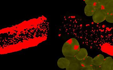
Photo by Ann Kuganathan
Niki Sadat Afjeh
Niki Sadat Afjeh
This image shows arteries—one with surrounding fat known as perivascular adipose tissue (PVAT), one without—from a spontaneously hypertensive rat (SHR). SHRs are a model used to study essential hypertension, which develops from an unknown cause and is exhibited by more than 95% of all hypertensive patients. The red staining on the vessels and the PVAT highlights oxidative stress, which is elevated in both tissues in hypertension. The vessels pictured are from a saline-treated SHR. However, the vessel on the right was co-incubated with PVAT from an SHR treated with an antioxidant protein called follistatin for 8 weeks. Upon a short 30-minute co-incubation, oxidative stress within the vessel was significantly reduced. Interestingly, chronic administration of follistatin also lowered blood pressure in these rats. PVAT-targeted therapy is a novel, growing field aimed at improving vessel health and thus, blood pressure.

Photo by Tran Minh Hai
Elham Sadat Abtahi
Elham Sadat Abtahi
Products made from fossil fuel derivatives have negatively impacted our global environment. Biodesign seeks to change that. Blue-green algae (cyanobacteria) can be found in abundance in lakes around the world. The remarkable fibre-like nets in this image were observed on the biomass of cyanobacteria through a focused microscopic lens. Cyanobacteria are the oldest form of life on earth with vibrant blue pigments. With this, we are developing a biological ink that will be more natural and safer than traditional inks often derived from heavy crude oil. The aim is to help reduce our carbon footprint on Mother Nature!

Photo by Annie DesRochers
This old pine stump, sitting on a rocky outcrop, bears witness to a distant past when the boreal forest reigned supreme. Through logging and fire, the black spruce disappeared and never returned. Why? We are in Rouyn-Noranda, close to the Horne smelter chimneys, which, up until 1970, spewed sulphuric and nitrous acid into the atmosphere. Hardly anything grows in this area, which is so acidic that even the rocks become brittle. Research is being carried out to reconstitute the soil and vegetation in order to bring life back to these desolate places.

Photo by Jacques Voisard
Since the 1950s, thanks to an electrode placed in the medial telencephalic bundle, we know that rats learn very quickly to operate a lever that stimulates the reward system in their brain. This bundle is a kind of highway through which the axons of countless neurons pass, to and from myriad zones in the brain. Which neurons are at work in the rewards system that we have been observing for so long? Only since the advent of the latest techniques in neural tracing has it been possible to separate the neurons that contribute to the reward system from those that do not.

Photo by Lisa Tischenko
Bits of DNA from the red oak tree float in these tubes like jellyfish. This chain-like molecule called DNA contains the genetic information in all living things—feathered, furry or leafy. From one individual to another, the information varies within a species according to their environmental conditions. The analysis of these samples will be used to identify genetic variation in populations of red oaks in northeastern America. In a context of climate change, it will be possible to select adapted seeds, favourable to the sustainable management of our forests.

Photo by Raphaël Dawant
These images of amazing but real colours show that it is possible to create relief on electronic circuits, usually in 2D. Adding in a few “valleys” paves the way for new electronic devices. The image on the left is made up of a single material (a polymer) and was created using lithography, a microfabrication technique; the colours show how nanometric thickness affects light reflection. The image on the right is made up of several materials, and is plasma etched: ions bombard the surface and create a highly controlled topography. The colours here depend on the metal used—ranging from blue for aluminum, to gold for titanium nitride.

Photo by Grégoire Bonnamour
This is a segment of sciatic nerve taken from a mouse. You can see, in red, the axon bundles that run along the nerve, surrounded by glial cells (green), which serve as their support. The glial cells have the capacity, thanks to the transcription factor Nr2f1 (blue), to differentiate into melanocytes, which colonize the skin and give it its colouring. In people with Waardenburg syndrome type IV, the expression of Nr2f1 is disrupted, resulting in a deficiency of melanocytes, and therefore a loss of pigmentation and balance. Melanocytes also play a key role in the workings of the inner ear!

Photo by Ophélie Martinie
The neuronal networks of our brain are like highways (yellow-orange) or secondary roads (blue). Here, seen from above by neuroimaging, are the two hemispheres of the brain of a 9-year-old child. Their asymmetry is striking. You can see the lesions that occurred during the development of the fetus, resulting in cerebral palsy. And, clearly visible, the left hemisphere contains far fewer roads. This results in motor problems, among other things. Deciphering the neural roadmap in affected children is crucial to better understand their symptoms and to improve interventions, which, to date, have proven only moderately effective.

Photo by Aida Romera Barbera
This tiny Trichuris trichiura egg is over 150 years old. It was found in the old latrine of a middle-class house from the beginning of the 19th century, in the heart of Old Limoilou in Quebec City. Although dead, the embryo is still visible in the center. Its discovery—a boon for archaeoparasitologists—confirms that at the time, when there were no sewage systems, many of our ancestors suffered from trichocephalosis, an intestinal parasitic disease caused by T. trichiura. There are an estimated 450 million to 1 billion active cases today, mostly in hot, humid countries with little infrastructure.

Photo by Redouane Amrar
Quantum computers will deliver phenomenal computing power compared to traditional computers. They will perform their calculations at the atomic scale, and for this, their microelectronic components such as memories and single-atom transistors will need to be miniaturized. To ensure the interconnection between these components, a process of evaporation of noble metal, e.g. gold (Au), is used. Even if this technique is well mastered, some surfaces may not always tolerate the deposition of the metal: as proof, check out these gold nanometric structures that have sprouted on a silicon chip and that evoke the tentacles of a sea anemone.

Photo by Walid Idi
This network of human neurons is observed not from a brain, but from a Petri dish. It was generated by the differentiation of neural progenitor cells. These neurons, which have the same characteristics as our own, have rapidly become mature and functional, allowing us to better understand, for example, the mechanisms of neurodegenerative diseases. This type of model can also be used in pharmacological and toxicological studies to evaluate the effects of drugs. Even if we are far from having a real brain, this neural network represents an excellent model for research!

Photo by Audrey Diouf-Lewis
Did you know that every year, Quebec farmers use 4000 tons of plastic film to wrap their hay bales? The COALIA team wants to give this single-use film a second life. As this microscopy image shows, the film is made of two different layers of plastic: polyethylene and EVA. This gives it flexibility and strength. Recycling, however, is complicated, as these plastics cannot be separated. However, the research team has succeeded in making new products, such as irrigation pipes for use in agriculture.

Photo by Claudia Covarrubias
Shown here strutting its ware is the detrusor, master of the urinary system. During urination, this muscle of the bladder contracts to evacuate the liquid. The study of these smooth muscle cells (in green) and their nuclei (in blue) allows us to identify the proteins involved in urinary incontinence associated with an overactive bladder. Ultimately, research on this muscle, whose function is regulated by the nervous system, may contribute to the treatment of other neurological conditions affecting urination, such as Parkinson's disease and multiple sclerosis.

Photo by Monsif Baraka
This is neither a wool sweater nor a stereogram (2D image giving the illusion of a 3D scene)! It is simply a corn field in the Bellechasse region. This image was taken at an altitude of 50 meters by a drone fitted with a multispectral camera. This advanced technique makes it easier to map crop residues, such as grass stalks, which makes it possible to determine the location and quantity of the residue. This data allows us to make better use of this plant material, which enriches the soil and cuts down on erosion. This is a great sustainable development practice.

Photo by Valérie Watters
For almost 15 years now, miniature and simplified versions of organs called organoids have been created in the lab. These three-dimensional structures are formed from stem cells that have the ability to differentiate into various types. Here we see a cross-section of a “mini-brain,” in which we can spot mature neurons in red, future neurons in green, and cell nuclei in blue. The doctoral student intends to verify how the addition of a long non-coding RNA (a molecule involved in genetic processes) can influence the formation of such a brain organoid.

Photo by Morgan Botrel
These floating islands are made up of tangled clusters of filamentous algae (chlorophytes) atop submerged aquatic plants. An unusual scene captured in Lake St. Pierre, a widening of the St. Lawrence River. Two weeks earlier, the islets were not even there. A rapid drop in the water level made them appear. Such a sudden appearance shows that aquatic grass beds are subject to extremely variable conditions. These habitats provide a variety of services, including the retention and removal of pollutants, including nitrates, the subject of this study. The image suggests that the purification capacity of such habitats depends more on the microscopic algae associated with the plants than on the plants themselves.

Photo by Ingrid Berenice Sanchez Carrillo
Although the nucleus of most cells is spherical or oval, there are exceptions. This image shows the epidermis of a leaf belonging to the species Arabidopsis thaliana, a model plant prized by biologists. After undergoing a genetic modification—the overexpression of a protein present in the nuclear pores—, the plant shows nuclei forming long filaments (in turquoise). In magenta, we see the chloroplasts responsible for capturing light during photosynthesis. To the researchers' surprise, this A. thaliana plant developed almost normally even though it was genetically modified. It remains to be seen what causes the deformation of the nuclei of the plant’s epidermis.

Photo by Alexis Leconte
This image of a mouse brain shows the tree-like structure of the animal's blood vessels. It was obtained without surgery, simply by placing a probe emitting ultrasounds on the rodent's head. Here we see the path of millions of microbubbles injected into the blood, which flow from the largest arteries to the smallest capillaries. We thus obtain images of the blood flow at an unprecedented level of detail! This technique is safe, as the microbubbles are rapidly eliminated by the lungs. In the long term, it could allow us to better understand the origin and progression of cerebral diseases such as Alzheimer's.